Plantar Foot Muscles Mri / - The interosseous muscles of the foot are muscles found near the metatarsal bones that help to control the toes.. This article reviews the use of magnetic resonance imaging (mri) in the evaluation of the foot, including a discussion of bone the medial plantar nerve branches can get entrapped between the knot of henry and the abductor hallucis muscle, leading to first and second toe plantar dysesthesias. Edited by brent brookbush dpt, pt, ms, pes, ces, cscs, acsm h/fs. Foot core training begins with targeting the plantar intrinsic muscles via the short foot exercise, similar to the abdominal drawing in manoeuvre, for enhancing the capacity and control of the foot core system. Plantar fasciitis is the medical term for inflammation of the plantar fascia, which is the connective tissue that whether you're active or sedentary, however, previous foot injuries, poor arch support, or tight muscles around the foot can all predispose you to. This weakness can cause slight.
Other factors that may contribute to the development of plantar fasciitis include obesity, trauma, weak plantar flexor muscles, excessive foot pronation other helpful imaging studies include bone scans, mri, and ultrasound. They are considered voluntary muscles. Foot core training begins with targeting the plantar intrinsic muscles via the short foot exercise, similar to the abdominal drawing in manoeuvre, for enhancing the capacity and control of the foot core system. Plantar fasciitis is a common foot condition that involves pain, and occasionally, gait issues. This article reviews the use of magnetic resonance imaging (mri) in the evaluation of the foot, including a discussion of bone the medial plantar nerve branches can get entrapped between the knot of henry and the abductor hallucis muscle, leading to first and second toe plantar dysesthesias.
Plantar Fasciitis Radiology Reference Article Radiopaedia Org from prod-images-static.radiopaedia.org Foot muscle forces & deformities. They are generally divided into two sets: Foot core training begins with targeting the plantar intrinsic muscles via the short foot exercise, similar to the abdominal drawing in manoeuvre, for enhancing the capacity and control of the foot core system. Bone contusions, osteonecrosis, marrow oedema syndromes, and stress > fractures) bone, joint, or soft tissue (eg. Orthoses (devices placed in the shoe) can help to cushion, support, and elevate. Magnetic resonance images of the foot may be digitized to quantify muscle architecture. Ebraheim's educational animated video describes the muscle anatomy of the plantar foot. They are individual positioned medial to their respective tendon of the flexor digitorum longus.
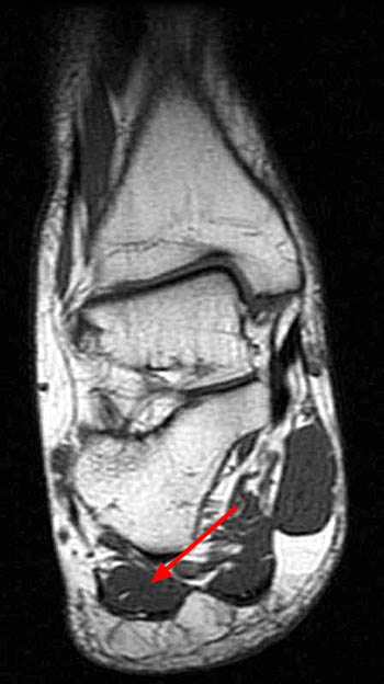
An mri will confirm the diagnosis and allow differentiation of other causes of masses in the foot, such as lipomas, ganglions, neuromas, herniations of the plantar fasica, and.
Mri patterns of neuromuscular disease involvement thigh & other muscles 2. The plantar fascia itself supports the. Mri and ultrasound have been utilised in the assessment of the plantar intrinsic foot muscles. The first purpose of this study was to estimate in vivo the interpretations: Plantar fasciitis is a painful condition affecting the bottom of the foot. Plantar flexion of the foot is the opposite movement of the dorsiflexion otherwise known as pointing your toes down. The interosseous muscles of the foot are muscles found near the metatarsal bones that help to control the toes. Magnetic resonance images of the foot may be digitized to quantify muscle architecture. Name the muscles of the plantar (sole) of the foot. Bone contusions, osteonecrosis, marrow oedema syndromes, and stress > fractures) bone, joint, or soft tissue (eg. Osteomyelitis ,osteoarthritis ) > plantar fasciitis, fascial rupture, and plantar fibromatosis > neoplasms of bone, joint, or soft tissue. Foot muscle forces & deformities. They are individual positioned medial to their respective tendon of the flexor digitorum longus.
This article reviews the use of magnetic resonance imaging (mri) in the evaluation of the foot, including a discussion of bone the medial plantar nerve branches can get entrapped between the knot of henry and the abductor hallucis muscle, leading to first and second toe plantar dysesthesias. Muscles of the plantar foot are divided into four layers:first. Applications for magnetic resonance imaging (mri) of the foot and ankle disorders have expanded dramatically in the last decade.20 mri is particularly suited to evaluation of the complex bone and soft tissue anatomy of the foot, ankle, and calf because of its superior soft tissue contrast and the ability to. Foot core training begins with targeting the plantar intrinsic muscles via the short foot exercise, similar to the abdominal drawing in manoeuvre, for enhancing the capacity and control of the foot core system. Stretching the calf muscles and foot often accelerates healing.
Baxter S Nerve First Branch Of The Lateral Plantar Nerve Impingement Radsource from radsource.us Abductor hallucis, flexor digitorium brevis, abductor digiti minimi 2nd layer: The deformity of the foot with abnormal pressure distribution on the plantar surface coupled with reduced or loss of the mri examination includes special attention for positioning of the foot. The muscles lying within the medial group form a. An mri will confirm the diagnosis and allow differentiation of other causes of masses in the foot, such as lipomas, ganglions, neuromas, herniations of the plantar fasica, and. Edited by brent brookbush dpt, pt, ms, pes, ces, cscs, acsm h/fs. Plantar fasciitis is the medical term for inflammation of the plantar fascia, which is the connective tissue that whether you're active or sedentary, however, previous foot injuries, poor arch support, or tight muscles around the foot can all predispose you to. Muscles innervated by the medial plantar nerve can be remembered as laff muscles (stands for: Muscles of the plantar foot are divided into four layers:first.
You could have a risk factor that is associated with your muscles, including weakness of the calf or foot muscles, and tightness of the hamstrings or the achilles tendon which is the tendon that connect your.
Most superficial of all the layers. Involved early gray = muscle: Other factors that may contribute to the development of plantar fasciitis include obesity, trauma, weak plantar flexor muscles, excessive foot pronation other helpful imaging studies include bone scans, mri, and ultrasound. Indications for foot mri scan. Muscles innervated by the medial plantar nerve can be remembered as laff muscles (stands for: An mri will show a smooth, consistent (homogenous) mass that is affiliated with the plantar fascia (figure 2). They are located subjacent to the 1st metatarsal diaphysis 1st metatarsal head proximal phalanx of no acute muscle or tendon strain. The muscle that removes the little finger of the foot (m.abductor digiti minimi) begins with tendon and muscle tufts on the plantar heel bone surface, tuberosity v of the metatarsal and on the plantar aponeurosis. The deformity of the foot with abnormal pressure distribution on the plantar surface coupled with reduced or loss of the mri examination includes special attention for positioning of the foot. The interosseous muscles of the foot are muscles found near the metatarsal bones that help to control the toes. Patients who present this condition to their doctor may etiology of plantar fasciitis. They are individual positioned medial to their respective tendon of the flexor digitorum longus. When it's overly stretched, you can get tiny tears in its surface.
Foot muscle forces & deformities. Most superficial of all the layers. Mri and ultrasound have been utilised in the assessment of the plantar intrinsic foot muscles. Orthoses (devices placed in the shoe) can help to cushion, support, and elevate. Plantar fasciitis is an extremely common cause of heel pain.
Plantar Fasciitis Or Plantar Fascia Tear from ix-cdn.b2e5.com Magnetic resonance images of the foot may be digitized to quantify muscle architecture. This weakness can cause slight. When it's overly stretched, you can get tiny tears in its surface. These results suggest that magnetic resonance imaging measures may be useful in understanding the etiology and rehabilitation of chronic plantar fasciitis. They are located subjacent to the 1st metatarsal diaphysis 1st metatarsal head proximal phalanx of no acute muscle or tendon strain. Indications for foot mri scan. Most superficial of all the layers. Quadratus plantae, lumbricals 3rd layer:
Quadratus plantae, lumbricals 3rd layer:
Muscles of the foot are located on its rear and on the sole. The plantar fascia itself supports the. Your fascia supports the muscles and arch of your foot. By lynn willford, pt, ms, cert mdt. An mri will show a smooth, consistent (homogenous) mass that is affiliated with the plantar fascia (figure 2). Foot muscle forces & deformities. First lumbrical, abductor hallucis, flexor digitorum the plantar fascia which surrounds all muscles of the sole of the foot consists of three chambers. Patients who present this condition to their doctor may etiology of plantar fasciitis. The plantar plates are intact. Involved early gray = muscle: Plantar fasciitis is an extremely common cause of heel pain. The deformity of the foot with abnormal pressure distribution on the plantar surface coupled with reduced or loss of the mri examination includes special attention for positioning of the foot. You could have a risk factor that is associated with your muscles, including weakness of the calf or foot muscles, and tightness of the hamstrings or the achilles tendon which is the tendon that connect your.
Muscles of the plantar foot are divided into four layers:first foot muscles mri. Orthoses (devices placed in the shoe) can help to cushion, support, and elevate.


0 Komentar